
Melanie Greenwood takes a trip to Bristol University’s School of Veterinary Science to see how this centre is becoming nationally-renowned for its high-tech treatments

Langford’s centre now handles around 2,000 equine cases from across the South West, South Wales, Gloucestershire, Wiltshire and Dorset annually.
Its latest addition is a new £700,000 diagnostic tool that has now arrived at the School of Veterinary Science.
The Hallmarq standing MRI (Magnetic Resonance Imaging scanner) is regarded as a major advance in imaging horses’ lower limbs and lameness diagnosis, which can affect an estimated 50 per cent of horses at some point.
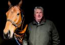
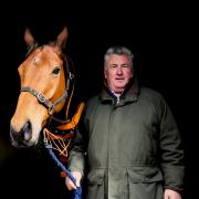
For four-year-old chestnut gelding, Pedran, the MRI scan is an option if an ultrasound can’t track down what’s really the matter. He is being petted and standing quietly sedated after getting his right back leg stuck in fence causing a painful swelling around his lower limb.
Veterinary Surgery Professor, Alistair Barr, says: “At around £1,000 for an MRI we may try other diagnostic tools first but sometimes an early accurate diagnosis and prognosis can save money and time in the long run and guide expectations more accurately.”
The equine scanner is housed within a copper-shielded trailer to prevent radio interference. It is temperature-controlled and once the horse is so deeply sedated it can stand motionless, it enters the trailer across a fluorescent yellow line.

The horseshoe-shaped magnet sits on the floor contained in royal blue padding and the horse is slowly coaxed into position and the process begins with results sent via computer.
Senior Equine Surgery Teaching Fellow, Evita Busschers, says: “The biggest advantage of the early use of an MRI is you know what you are treating before it has the potential to become a disaster.”
As well as lameness many horses are referred to Langford because of head and jaw injuries or sinus and dental problems.
For this the school’s £250,000 CAT (Computerized Axial Tomography) scan is used to take data from a mass of revolving X-ray images, which are converted into three-dimensional highly-detailed pictures.
Senior Equine Surgery Lecturer, Henry Tremaine, says:“We’ve seen a number of jaw fractures from the restraint of a horse when it’s suddenly panicked and pulled or where it’s been badly kicked by another horse.
“Once sedated, the horse steps on a platform supported by a cushion of air which is effortless for us to move and means its head can be carefully placed inside the CAT scanner. It takes just 45 seconds and is an amazing help pre-surgery.”

The ABC of MRI scans
MRI stands for Magnetic Resonance Imaging and it uses exceptionally strong magnet and radio frequency waves to reveal complex, hidden, internal images.
The first full body scan to diagnose cancer in humans was performed in 1977 and was the brainchild of American medical researcher Raymond Vahan Damadian who discovered that cancer tumours and normal body tissue differed in response to magnetic exposure.The human body is made up of water molecules - hydrogen and oxygen atoms. At the centre of each hydrogen atom there is an even smaller particle called a proton. Protons are like minute magnets which are hypersensitive to magnetic fields. The magnet forces these tiny protons to line up in the same direction just as a magnet pulls at the needle in a compass. Short bursts of radio waves are then pulsed into the leg, or hoof, disturbing the protons out of their magnetic alignment. When the radio waves are switched off the protons naturally realign sending out their own faint radio signals which are analysed by computers. Because protons realign with different speeds and distinct signals the picture they create provides in-depth information about what is going on deep within the skin.
Equine lameness
Lameness is an abnormal gait or stance and it’s thought as many as 50 per cent of owners deal with this ailment at some point. It is the most common and costly problem in horses in terms of diagnosis and treatment. Much lameness in horses arises within the feet and an MRI scan is regarded as a gold standard for diagnosis as it offers exceptional evaluation of both soft tissue and bone change. Prior to MRI scans, examinations are carried out by X-ray and ultrasound, however these provide much less detail, particularly for soft tissue injuries within the foot. The MRI improves the accuracy of diagnosis and prognosis and helps plan appropriate treatment and rehabilitation so owners are more realistic about the horse’s future health. Many degenerative conditions like osteoarthritis and chronic tendon and ligament injuries remain unlikely to be permanently cured and are challenging to manage, however accurate assessment allows all concerned to work from a better informed starting point.